Neuro Discovery Platforms
Need to explore mechanism of action to select a better candidate to progress into in vivo studies? Our NerveSim® and BrainSim® are the only models in the industry that provide high levels of physiologically-relevant myelination and we use the same metrics used by clinicians to diagnose and track disease progression, nerve conduction velocity, and histomorphometry. Our models provide clinically predictive data faster, helping take the guess work out of candidate selection. We also offer 2D mono- and co-culture systems to assist with candidate selection if you are still in the process of narrowing your pipeline.

Need a predictive model of peripheral neuropathy? Need clinically relevant data? Our NerveSim® 3D in vitro peripheral nervous system model can provide answers to your safety and efficacy questions.
Our NerveSim® platform is the only in vitro model with Schwann cell myelination and human-relevant electrophysiological and structural data metrics, unlocking a powerful tool for human translation. In addition, we offer rodent models to assist with bridging the gap between historical in vivo data to in vitro and ultimately to human relevant data.
We grow our neuronal tissue in a proprietary 3D environment to mimic nerves in both form and function. NerveSim® outputs the most clinically-relevant readouts for peripheral neuropathies and neuropathic pain: nerve excitability, conduction, and histomorphometry.
PLATFORM STEPS
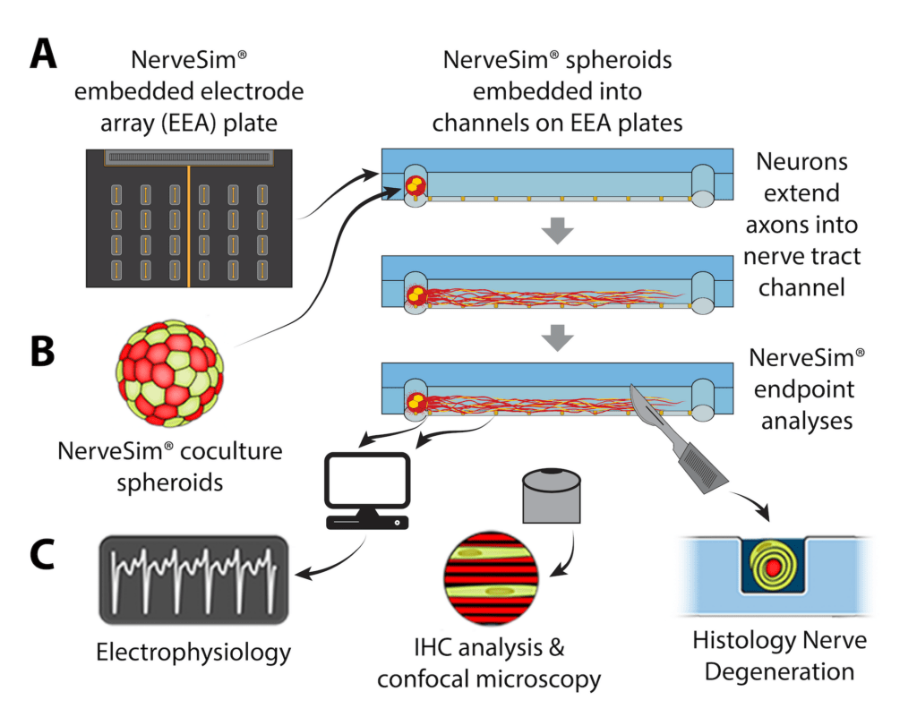
We first create the building blocks for the platform by organizing multi-cellular primary cultures, or iPSCs, into 3D organoids, seen on the left as either rodent of human spheroids. Next, we implant the spheroids into our NerveSim® to grow biomimetic tissue, for an unparalleled model of the peripheral nervous system. This design allows for the same testing that is done in the clinic and in vivo, but in vitro.
After culturing and growing each construct, we assess the electrophysiology to measure clinically-relevant changes in neuronal-glial properties reflective of the effects on the peripheral nervous system in response to insult. Our nerve conduction velocity (NCV) and nerve excitability measurements demonstrate a more sensitive system for detecting neurotoxicity, and subsequent rescue with therapeutics, when compared to 2D assays. Until now, this metric was only available through in vivo testing.
By correlating structural changes to functional metrics through histomorphometry, we gain unparalleled insights into the mechanism of action that would normally take millions of dollars and several years of testing in addition to clinical trials to achieve. This is how we provide human data, faster to our clients and partners.

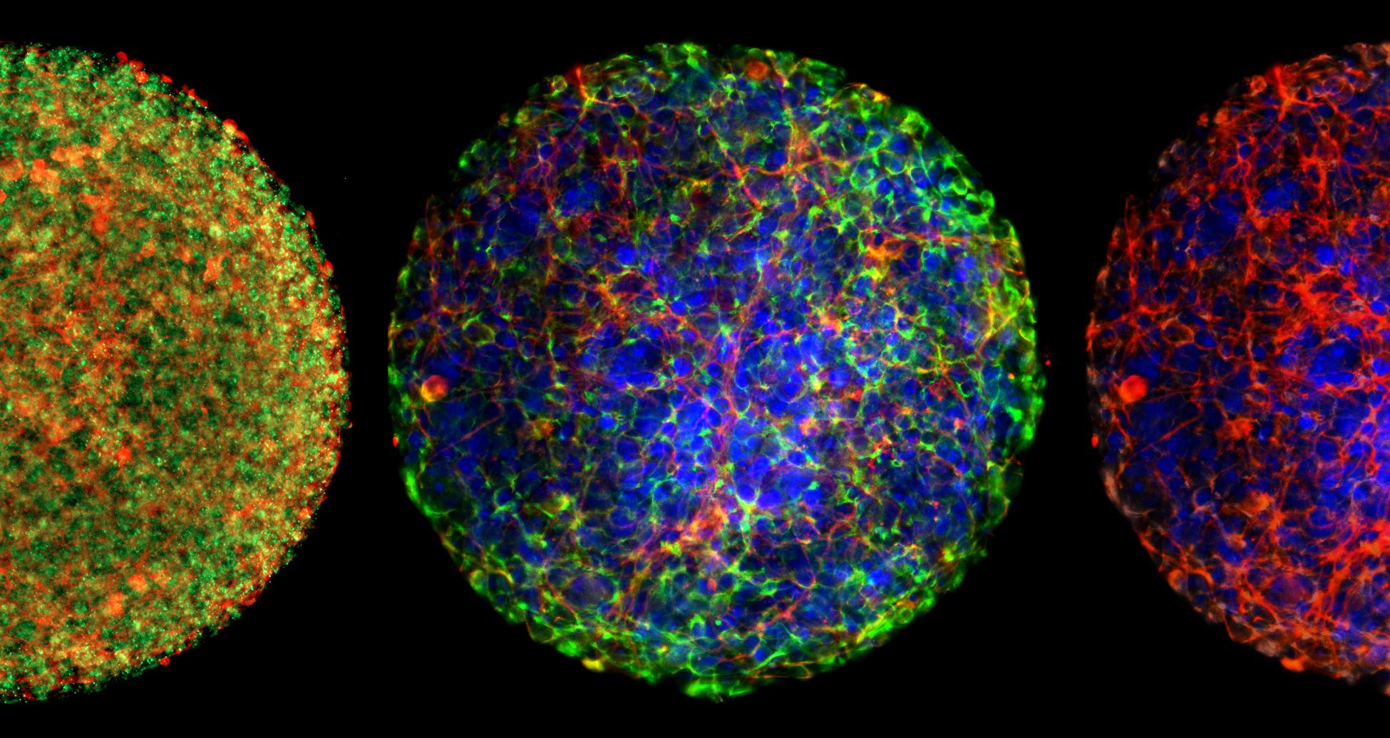
AxoSim’s BrainSim® technology enables human-relevant oligodendrocyte myelination, representing a breakthrough for testing in neurotoxicity and neurodegenerative diseases. This proprietary platform was invented in 2016 by Dr. Thomas Hartung at Johns Hopkins and has been exclusively licensed by AxoSim. Our BrainSim® platform recreates a truly biomimetic 3D environment, featuring a unique combination of neurons, astrocytes, and oligodendrocytes.
We begin by differentiating iPSCs into NPCs to create the building blocks for a multi-cellular biomimetic platform. This proprietary differentiation technique produces three critical cell types: neurons, astrocytes, and oligodendrocytes in our BrainSim® spheroids. To ensure quality and reproducibility we maintain precise control of spheroid size to avoid necrotic cores. Our methods of culturing these spheroids influences cell to cell interaction and induce high levels of myelination.
In addition, our BrainSim® replicates critical characteristics of neurons and glia to create a better representation of the brain’s structure. We then use this model to characterize cellular responses through electrophysiology, ICC, histology, flow cytometry, and gene expression and can correlate phenotypic changes to a drug’s mechanism of action, providing meaningful data faster and more accurately than previously possible.
The microBrain™ technology provides a physiologically-relevant human iPSC-derived screening platform for CNS drug discovery and toxicity testing. The organoid platform replicates important human CNS biology, containing cortical neurons and astrocytes that self-organize and form functional networks in 3D. The hallmark of the microBrain organoids, are their remarkably consistent performance, making it ideal for high throughput neurotoxicology, neuromodulation, and drug discovery investigations.
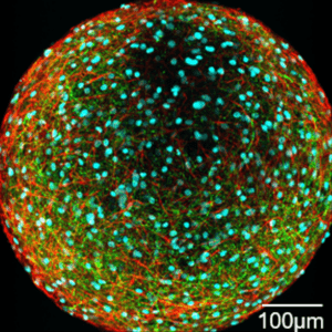 Neural progenitor cells (NPCs) are seeded into ultra low attachment (ULA) plates where they self-organize into 3D spheroids. Over the course of 6 weeks, NPCs co-differentiate into neurons and astrocytes. Differentiated microBrain organoids exhibit spontaneous and coordinated network activity.